Positron emission tomography (PET) scanning technology has now been in place at the UC Davis veterinary hospital for two years. In 2016, UC Davis became the first veterinary hospital in the world to implement an equine PET scanner.
The program has enjoyed a successful second year with many exciting developments.

Through collaborative support from the UC Davis Center for Equine Health and Brain Biosciences, specialists with the Diagnostic Imaging Service have now completed more than 85 equine PET studies. These include important research projects and the growth of clinical purposes for PET scans to improve equine health through the utilization of the piPET device, a scanner originally developed for neuroimaging studies in non-human primates but found by UC Davis researchers to be quite useful on horse legs.
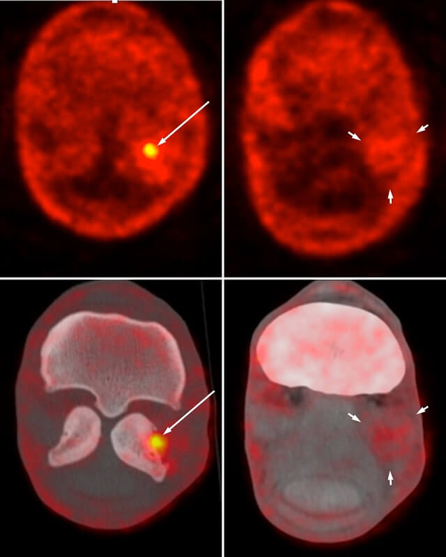
One of this year’s most important achievements was the validation of a dual-tracer scanning protocol, which allows for the assessment of both bone and soft tissue lesions in lame horses. This protocol has now been used on several clinical cases and has been utilized to assess complex lesions involving the foot and the suspensory ligament. (Figure 1)
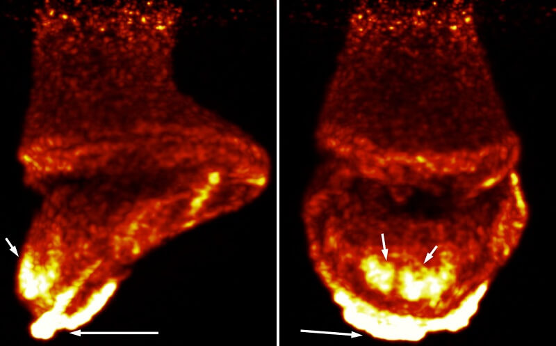
PET has also proven to be an interesting component for laminitis research. Laminitis, as most horse owners know, is an extremely serious condition of the horse foot that, unfortunately, is often fatal. There are still a lot of unknowns on the origin and development of the disease. While researchers continue to investigate the disease, PET has brought new insights on the areas of the foot that are the most affected. (Figure 2)

PET also contributed to research in regenerative medicine at UC Davis. Working with veterinarians in the Equine Lameness and Surgery Service, radiologists have been able to label stem cells with PET tracers, allowing to assess their distribution after injection and confirming their location in targeted lesions. Radiologist Dr. Mathieu Spriet had extensive experience with tracking stem cell using scintigraphy and has been quite impressed with the advantages that PET brought.
“With PET tracking, we are now able to confirm the presence of stem cells in a specific lesion in a tendon,” said Dr. Spriet. “Using scintigraphy, we were limited to only being able to tell that stem cells were present in the region of the foot.”
In addition to multitude of research studies, UC Davis has scanned more than 40 clinical patients, bringing useful information for management of cases. In one case, Irish Streetsinger—a racehorse with persistent lameness—was admitted to UC Davis. Following several lameness evaluations (including radiographs and nuclear scintigraphy to determine the area of lameness), clinicians advised her owner that a PET scan would be the best option to obtain a conclusive diagnosis. The scan pinpointed her lameness, and following stem cell treatment, she was back on the track. In 2018, Irish Streetsinger ran seven races with one victory.
Drs. Spriet and Pablo Espinosa have presented their PET research at multiple regional, national and international conferences, including annual meetings of the American College of Veterinary Radiology, the European College of Veterinary Diagnostic Imaging, and the Society of Nuclear Medicine and Molecular Imaging.
“We are seeing a tremendous amount of enthusiasm for the use of PET from the veterinary imaging community,” said Dr. Spriet. “Members of veterinary imaging organizations from around the world are excited to learn about our advancements for the capabilities of equine PET imaging.”

News from the horse industry. Sharing today’s information as it happens. The Northwest Horse Source is not responsible for the content of 3rd party submissions.


